
- QUASAR
- AquaCycle
- Proteins4Singapore
- Singapore's Pathway to Carbon Neutrality
- CellFACE
- LightSPAN
- Computational Modelling Group
- Energy and Power Systems Group
- SITEM - Singapore Integrated Transport and Energy Model
- Past Projects
CellFACE
Executive Summary
CellFACE aims to establish imaging flow cytometry as a label-free platform technology for routine cellular diagnostics at the point-of-care. To assure success of the interdisciplinary CellFACE project, back-to-back teams between Munich and Singapore are formed. Using co-creational principles engineers and life scientists at TUM-CREATE, NTU, and A*STAR cooperate with clinicians at National University Hospital to develop a workflow solution for blood cell analysis to improve acute care diagnostics.
Research
Clinical and diagnostic challenges
Approximately 5% of all adult presentations and up to 15% of visits from elderly patients to emergency rooms (ER) are for fever [1]. Clinicians face several dilemmas because it is the initial symptom presenting for a range of illnesses, from a simple viral infection to meningitis or malignancy. For instance, in Singapore fever constituted 21.6% of all unscheduled patient return visits to the ER of which 28.3% had a wrong or delayed diagnosis [2] - in particular elderly patients [3-5]. Depending on the complexity of the case, inadequate patient management leads to extra costs and ineffective allocation of clinical resources at ERs, the intensive care/acute medical units (ICUs/AMUs), and wards. Time-to-results for a correct differential diagnosis for the cause of fever adds on average of 10.000 S$ for in vitro diagnostics (IVD), medical imaging per hospitalized NUH patient, and unnecessary application of antibiotics.
Blood cells are of highest importance as biomarkers in the clinical routine due the unique properties of cellular biomarkers. Today, automated hematological analysis is in fact one of the most requested in vitro diagnostic tests worldwide providing complete blood counts (CBC) as well as leukocyte differentials (Diff). Despite the availability of automated analyzers in the central laboratory, the gold standard for the routine diagnosis of hematological disorders is the tedious Giemsa stained blood smear and is always required when analyzer report a flagged result. In the central laboratory, flagged results are a break of automation and are cost drivers. The analysis of thin blood films require personnel, suffer from inter-observer variations due to low statistical power. Most importantly, a CBC/Diff only provides information on blood cell concentrations but lacks functional cell information.
Scientific and technical goals
Imaging flow cytometry based on a highly defined microfluidic flow of blood samples and digital holographic microscopy (DHM) allows to perform high-throughput cell analysis at physiological conditions. Due to the label- and reagent-free imaging approach we can visualize cell morphologies, differentiate cells based on phase information [6] and - most importantly - have access to hidden functional biomarker. The value of these new classes of biomarkers will be observed in longitudinal clinical studies at NUHS. In this way, we hope to support rapid stratification of patients with fever using functional hematology biomarkers related to immune system, hemostasis, and organ function.
Cartridge-based workflow integration with industry partner and artificial intelligence algorithm will be explored to reduce workflow complexity and computational effort. Robust analysis of hidden biomarker will demonstrate the realization potential of future point-of-care (POC) applications with time-to-results in a few minutes. To ensure reproducible clinical testing, CellFACE will develop additional standardization and calibration methods for this next generation POC hematology analyzer. The results of CellFACE could potentially revolutionize patient care, producing many benefits ranging from improved antimicrobial stewardship to prevention of unnecessary hospital admissions during uncertainty of diagnosis, and help save costs by focusing investigations to rapidly identify the underlying causes of fever.
.JPG)
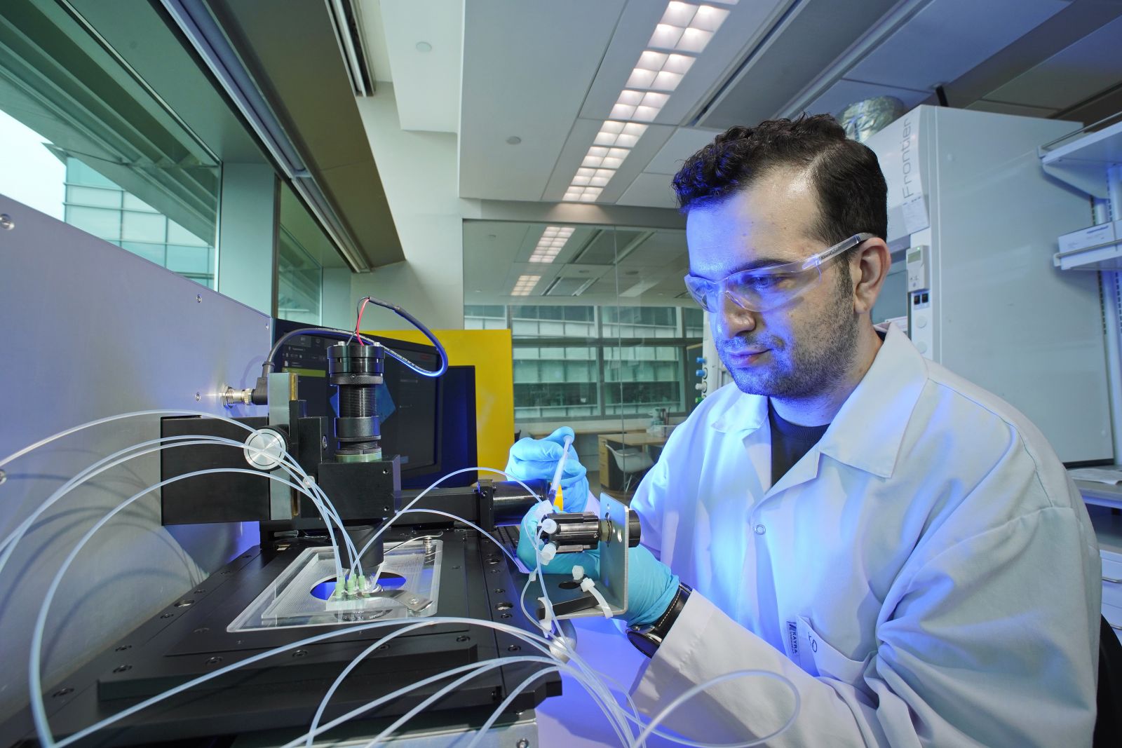
Figure 1. Microfluidic channel for highly parallelized blood cell imaging (© Richard Koh) Figure 2. CellFACE prototype operated in a Biosafety Level 2 laboratory at CREATE (© Richard Koh
Members
Organization Name Designation
TUM
Diepold, Klaus
PI, Professor
NA
Hayden, Oliver
PI, Professor
NA
Knolle, Percy
PI, Professor
NA
Prazeres da Costa, Clarissa
PI, Professor
NA
Schneider, Gerhard
PI, Professor
NA
Hamway, Youssef
Postdoc
NA
Irl, Hedwig
Physician
NA
Klenk, Christian
PhD
NA
Röhrl, Stefan
PhD
TUMCREATE
Delikoyun, Kerem
Research AssociateChen, Qianyu Research Associate Si Ko Myo Quality Software Engineer
A*Star
Renia, Laurent
PI, Professor
NA
Lee, Hwee Kuan
PI, Adjunct Associate Professor
NA
Wei, Liu
Postdoc
NA
Wang Wei, Charles
Supplier
NTU
Hou, Han Wei
Assistant Professor
NUHS
Chew, Ka Lip
PI
NA
Chng, Wee Joo
PI, Professor
NA
Cove, Matthew Edward
PI, Assistant Professor
NA
Kuan, Win Sen
PI, Assistant Professor
NA
Soong, John Tshon Yit
PI, Assistant Professor
Sunningdale Tech Ltd
Goh, Yew Guan
Industry Collaborator
Dracoon GmbH
Schieder, Marc
Industry Collaborator
Duke-NUS
Bertoletti, Antonio
AdviserContact
For any enquiry on CellFACE project, please contact us at:
Email: cellface@tum-create.edu.sg
Phone: +65 9784 9125
Publications
[1]
S. DeWitt, S. A. Chavez, J. Perkins, B. Long, and A. Koyfman, “Evaluation of fever in the emergency department,” Am. J. Emerg. Med., vol. 35, no. 11, pp. 1755–1758, Nov. 2017, DOI: 10.1016/j.ajem.2017.08.030.
[2]
C. H. W. Soh et al., “Risk Factors for Emergency Department Unscheduled Return Visits,” Medicina, vol. 55, no. 8, p. 457, Aug. 2019, DOI: 10.3390/medicina55080457.
[3]
G. Gavazzi and K.-H. Krause, “Ageing and infection,” The Lancet Infectious Diseases, vol. 2, no. 11, pp. 659–666, Nov. 2002, DOI: 10.1016/S1473-3099(02)00437-1.
[4]
K. P. High et al., “Clinical Practice Guideline for the Evaluation of Fever and Infection in Older Adult Residents of Long-Term Care Facilities: 2008 Update by the Infectious Diseases Society of America,” Clinical Infectious Diseases, vol. 48, no. 2, pp. 149–171, Jan. 2009, DOI: 10.1086/595683.
[5]
M. A. Horan and N. Pendleton, “The relationship between aging and disease,” Rev. Clin. Gerontol., vol. 5, no. 2, pp. 125–141, May 1995, DOI: 10.1017/S0959259800004093.
[6]
M. Ugele et al., “Label-Free High-Throughput Leukemia Detection by Holographic Microscopy,” Adv. Sci., vol. 5, no. 12, p. 1800761, Dec. 2018, DOI: 10.1002/advs.201800761.
Team
Dr. Kerem Delikoyun Research Fellow Qianyu Chen Research Associate